The Decades-Old Mystery Inside Your Ultrasound Machine—Solved by a 3D Atomic Map
CAMBRIDGE, MASS. — May 2026 – For decades, a class of materials called relaxor ferroelectrics has powered technologies that touch nearly every human life. Ultrasound machines that image unborn children. Sonar systems that navigate submarines. Precision microphones that capture sound. Actuators that move with nanometer precision. These materials are everywhere, hidden inside devices that work so reliably that no one thinks to ask how.
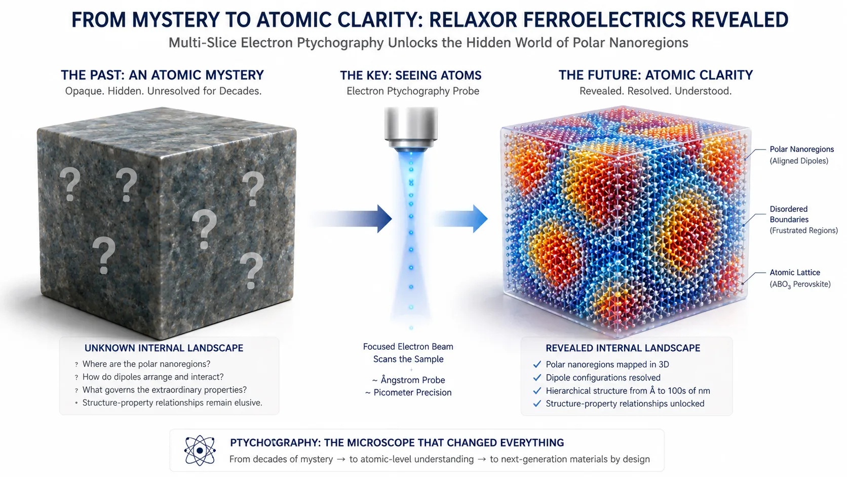
The answer, it turns out, is that no one fully knew. Relaxor ferroelectrics derive their extraordinary properties from their internal atomic structure—a delicate, nanoscale arrangement of electric charges that responds to external fields with remarkable efficiency. But that structure has stubbornly eluded direct measurement. The leading simulations suggested a certain organization of polar nanoregions—tiny zones where electric dipoles align—but no instrument had ever been able to see them in three dimensions. The models were hypotheses. The reality was invisible.
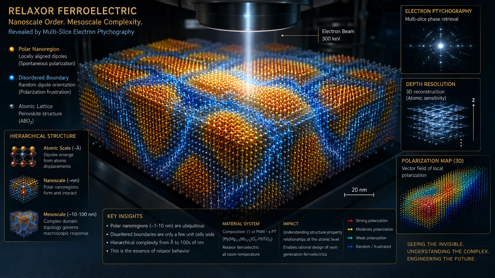
In April 2026, a team from MIT and collaborating institutions made it visible. Published in Science, their study provides the first direct three-dimensional atomic-scale characterization of a relaxor ferroelectric. Using a cutting-edge technique called multi-slice electron ptychography, the researchers mapped the distribution of electric charges across the material with unprecedented resolution. What they found was not what the models predicted.
The Ptychography Revolution
The technique that made the discovery possible is itself a story of scientific persistence. Multi-slice electron ptychography, or MEP, involves moving a nanoscale probe of high-energy electrons over a material in a precise grid pattern. At each position, the instrument measures the resulting electron diffraction pattern—the way the electron beam scatters as it passes through the atomic structure. Because each measurement overlaps slightly with its neighbors, powerful algorithms can reconstruct the three-dimensional arrangement of atoms and their associated electric fields from the diffraction data.
"It's like doing a CT scan, but at the atomic scale," explained co-first author Michael Xu. "We collect thousands of diffraction patterns, each containing information about the material's structure at that specific location. By combining them computationally, we can reconstruct the full three-dimensional picture."
The technique revealed a hierarchy of chemical and polar structures spanning from atomic to mesoscopic scales—structures that had been hypothesized but never directly observed. Most significantly, the researchers found that many of the polar nanoregions in the material were much smaller than the leading simulations had predicted. The models, it turned out, were incomplete. The reality was more complex, more heterogeneous, and more beautiful.
Why It Matters
The significance of the MIT team's achievement extends far beyond academic satisfaction. Relaxor ferroelectrics are critical materials for energy storage, sensing, and electromechanical conversion. Their ability to store and release electrical energy efficiently makes them candidates for next-generation capacitors in electric vehicles and power grids. Their sensitivity to mechanical deformation makes them ideal for the piezoelectric sensors in medical imaging and industrial monitoring. And their unique electromechanical coupling properties are being explored for solid-state cooling applications.
But engineering these materials—tuning their composition and processing to achieve specific performance targets—has been an exercise in trial and error, guided by models that were known to be incomplete. With the 3D atomic structure now directly measured, those models can be refined. The MIT team fed their experimental data into simulations developed by collaborators at the University of Pennsylvania and the University of Alabama at Birmingham, creating a feedback loop between measurement and theory that will accelerate materials design.
"This study is the first time in the electron microscope that we've been able to directly connect the three-dimensional polar structure of relaxor ferroelectrics with molecular dynamics calculations," Xu said. "We can now see what the simulations need to reproduce, and we can validate whether a given model is capturing the real physics."
The implications for product design are substantial. A medical device company developing a next-generation ultrasound transducer, for example, can now work with materials models that have been validated against direct atomic-scale observation rather than inferred from bulk property measurements. The design cycle can accelerate, and the performance targets can be more ambitious, because the underlying physics is better understood.
What Every Entrepreneur Can Learn
The relaxor ferroelectric breakthrough offers lessons that transfer well beyond materials science.
First, measurement enables design. The history of technology is the history of better instruments enabling better products. The steam engine preceded thermodynamics. The transistor preceded solid-state physics. But in each case, the transition from trial-and-error to theory-driven design came only after the underlying phenomena could be measured. The MIT team's 3D atomic map of relaxor ferroelectrics is a measurement that will enable design. Entrepreneurs in any science-based industry should ask: what fundamental measurement has not yet been made, and who would benefit if it were?
Second, the gap between simulation and experiment is a market opportunity. The leading models of relaxor ferroelectrics were good enough to guide research but not accurate enough to replace physical testing. The MIT team's data provides a benchmark against which simulations can be validated, narrowing the gap. In any field where simulation and experiment diverge, there is value in closing the gap—through better measurement, better modeling, or both.
Third, decades-old mysteries are often hiding in plain sight. Relaxor ferroelectrics have been in commercial use since the 1980s. Ultrasound machines, sonar systems, and precision actuators have been built and sold for generations. And yet, until April 2026, no one had ever seen the atomic structure that gives those devices their capabilities. The most valuable scientific and commercial discoveries are often not new materials, but new understanding of materials we already use.
The Road Ahead
The MIT team's work is not the end of the story. It is a foundation. With the 3D atomic structure now known, materials scientists can begin the systematic work of engineering relaxor ferroelectrics for specific applications—tuning the size and distribution of polar nanoregions, optimizing the chemical composition, controlling the processing conditions to achieve desired performance. The models will improve. The design tools will become more powerful. The materials will get better.
For the industries that depend on relaxor ferroelectrics—medical imaging, defense, industrial sensing, energy storage—the implications are incremental but cumulative. Better materials enable better devices. Better devices enable better outcomes. The ultrasound machine of 2035 will not look dramatically different from the ultrasound machine of 2025. But the material inside its transducer will be smarter, more efficient, and better understood, because a team at MIT finally saw what had been invisible for four decades.